112年:醫學五(1)
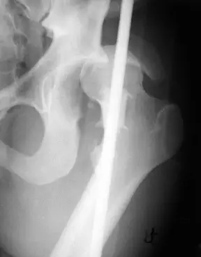
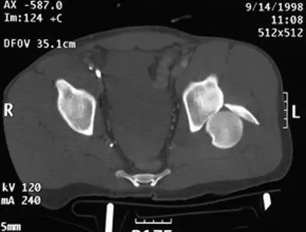
20歲男性病人因車禍左髖關節腫痛。圖為本次就診之左髖關節 X 光攝影及電腦斷層影像。最可能的診斷為何?
Aanterior hip fracture-dislocation
Bposterior hip fracture-dislocation
Cheterotopic ossification following dislocation
Davascular necrosis of femoral head
詳細解析
本題觀念:
本題核心在於鑑別髖關節脫位的方向與合併骨折,特別是後方髖關節骨折脫位(posterior hip fracture-dislocation)的影像學特徵與臨床意義。
影像分析:
從骨盆正位X光片可見左側股骨頭與 acetabulum 失去正常對位關係,股骨頭相對於右側明顯向上、向後且輕微外隱,股骨小轉子在AP投影中不明顯,暗示內旋位置。左側股骨頭尺吋於幾何放大下比右側小,符合股骨頭後方移位的特徵(radiopaedia.org)。
進一步於 axial CT 平掃中,可見左股骨頭完全移出髖臼,位於 posterior acetabular rim 之外,且緊鄰一塊骨片,代表 posterior rim fracture。周邊尚見關節腔內軟組織密度,符合急性髖關節骨折脫位的表現(radiopaedia.org)。
選項分析
- 選項A(anterior hip fracture-dislocatio
...(解析預覽)...
![醫師[2] - 下肢與軸性骨折 - AI 圖文解析預覽](/_next/image?url=https%3A%2F%2Fbgvxfcrmbdvefjhuvrmt.supabase.co%2Fstorage%2Fv1%2Fobject%2Fpublic%2Fvisual-explanations%2F192%2F7684_tb10bd3ed.webp&w=1920&q=75)
升級 VIP 解鎖圖文解析