107年:核醫診療(2)
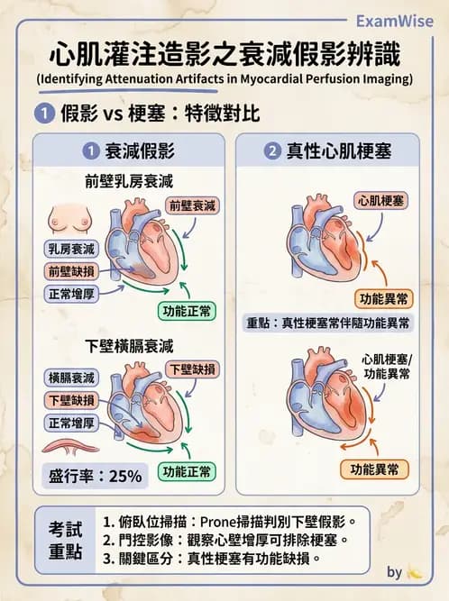
在門控心肌灌注造影的影像上若看到「左心室下壁」有固定性灌注缺損( fixed perfusion defect ),但門控功能分析上該區域卻呈現正常的心壁運動( wall motion )與增厚( wall thickening ),這種缺損應該是下列何種假影所造成?
A乳房衰減假影( breast attenuation artifact )
B橫膈衰減假影( diaphragm attenuation artifact )
C移動假影( motion artifact )
D散射假影( scatter artifact )
詳細解析
本題觀念:
本題考察門控心肌灌注造影(gated myocardial perfusion imaging)中,**固定性灌注缺損(fixed perfusion defect)伴隨正常心壁運動(wall motion)與增厚(wall thickening)**的臨床意義,此組合是衰減假影的典型表現,尤其針對「左心室下壁」的缺損,需鑑別橫膈衰減假影(diaphragm attenuation artifact)。
選項分析
(A) 乳房衰減假影(breast attenuation artifact) 此選項錯誤。乳房衰減假影通常出現在前壁(anterior wall)或前側壁(anterolateral wall),是由乳房組織對前方 gamma 射線的衰減所致,好發於女性受檢者。題目描述的缺損位於**「左心室下壁」**,不符合乳房衰減的典型位置。
(B) 橫膈衰減假影(diaphragm attenuation artifact) 此選項正確。橫膈衰減假影是左心室下壁(inferior wall)最常見的衰減假影來源,典型特徵為:
- 位置:左心室下壁(inferior wall)
- 灌注影像:固定性缺損(在壓力和靜息影像上均可見)
- 門控功能分析:心壁運動(wall motion)與增
...(解析預覽)...
升級 VIP 解鎖圖文解析